- Who we are
- Schools and Training
- Agenda
- Events
- Patients
- Payments
- FAQ
- Y-SICCR

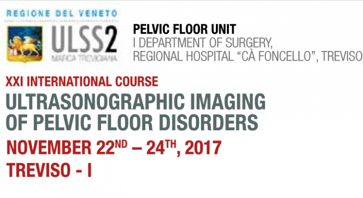
Imaging is gaining a key role in the understanding of pelvic fl oor disorders. Three-dimensional endoanal, endorectal and endovaginal ultrasonography, and dynamic transperineal US (DTPUS) are nowadays increasingly used in clinical practice for patients suff ering from pelvic floor dysfunctions (fecal and urinary incontinence, pelvic organ prolapse, obstructed defecation) and for patients with anal and rectal pathologies, either benign (anorectal sepsis) or malignant (anorectal tumors). These noninvasive techniques not only provide a superior depiction of the pelvic anatomy but also yield unique dynamic information. The new 3D technology is on the front line in the diagnostic and therapeutic workup and is superior to 2D-US, due to the elaboration of images, achieving better accuracy in the diagnosis of complex diseases.
This course is organised for all physicians who are involved or interested in pelvic fl oor disorders: general surgeons, gastroenterologists, radiologists, urologists and gynaecologists. Theoretical session presents the main guidelines and indications for the use of the different US techniques in clinical practice, demonstrating the endosonographic appearance of normal anatomy and common pathologic conditions of the pelvic floor. Practical session is organised in order to provide the participants with hands-on experience in the basic use of US and is designed to closely discuss specific details of the techniques.
Finally the course includes a session of hands-on training on PC-3D view workstation to let each participant review, manipulate, analyze and discuss several clinical cases under experts’ advice. A number of highly regarded specialists in this area have been selected to provide their experiences on thedifferent topics of the course.
COURSE OBJJECTIVES
• To cover a broad spectrum of ultrasonic techniques in pelvic floor disorders
and to provide the participants with didactic education in the indications for the use of ultrasonographic imaging of pelvic floor disorders in
clinical practice
• To demonstrate the endosonographic appearance of normal anatomy and common benign and malignant pathologic conditions of the pevic
organs
• To provide hands-on sessions to highlight technicalities of ultrasound through the broadcasting of live procedures, to allow real-time discussion between the examiners and the trainees and to improve skills of participants in ultrasound through practice on live or laptop computers under experts’ tutorials
PRESENTATION FACULTY
R. BACCICHET Treviso-I M. BARBAN Treviso-I
H. HERAS Treviso-I
A. INFANTINO S.Vito al Tagliamento-I
A. LAURETTA S.Vito al Tagliamento-I
G. MORANA Treviso-I F. MUGGIA Treviso-I
C. PASTORE Treviso-I
G. A. SANTORO Treviso-I
P. WIECZOREK
SCIENTIFIC SECRETARIAT
G.A. SANTORO
Head, Pelvic Floor Unit Treviso – Italy
giulioasantoro@yahoo.com
Pelvic Floor Unit, I Department of Surgery,
Regional Hospital “Cà Foncello”, Treviso – Italy
keycongress&communication
Via Makallè, 75 – 35138 Padova
Tel. ++39 049 8729511 Fax ++39 049 8729512
www.keycongress.com registration@keycongress.com
Last insertion:
Toilet Training
Read ›
Dott.ssa Paola De Nardi
Rubrica diretta a quanti volessero porre dei quesiti al Presidente SICCR, Dott.ssa Paola De Nardi.